
Introduction to Modern Medical Imaging
In July 2026, medical imaging stands as a cornerstone of modern healthcare. These advanced technologies allow us to peer inside the human body like never before. They are vital for diagnosing diseases early and accurately. This extensive guide explores the incredible innovations that have shaped medical imaging. We will look at how these advancements improve diagnostic accuracy and patient outcomes. We will also consider the future role of radiologist education in this fast-evolving field.
Evolution of Diagnostic Medical Imaging
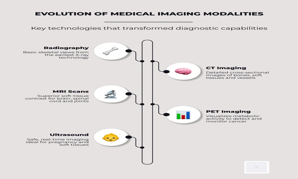
From the first rudimentary X-rays, medical imaging has undergone a remarkable transformation. Early radiography gave us basic skeletal views. This was a start. Over time, technologies like computed tomography (CT) and magnetic resonance imaging (MRI) emerged. Nuclear medicine and ultrasound technology also became essential tools. Each new modality dramatically expanded our diagnostic capabilities. These developments have progressively enhanced our understanding of health and disease.
Clinical Applications of Sub-Specialized Medical Imaging
Today, medical imaging is indispensable across almost all medical specialties. For instance, CT scans are crucial for trauma assessment and cancer staging. MRI excels in diagnosing soft-tissue and neurological disorders. Positron Emission Tomography (PET) is key for metabolic and oncologic imaging. We rely on these techniques for everything from oncology staging to cardiovascular assessment. They are vital for orthopedic evaluation and for women’s health initiatives such as 3D mammography. The ability to perform Sub-specialized medical imaging enables highly targeted, precise diagnoses, ensuring patients receive the most accurate care.
Here are key clinical applications for major imaging modalities:
Advanced Modalities and Hybrid Technologies
The quest for ever-greater diagnostic precision has led to the development of advanced modalities and hybrid imaging systems. These systems combine the strengths of different technologies, offering a more comprehensive view of the body’s anatomy and function.
Hybrid systems like PET/CT, SPECT/CT, and PET/MRI are at the forefront of this innovation. PET/CT, for example, merges metabolic information from PET with anatomical detail from CT, providing precise localization of disease activity. Similarly, SPECT/CT combines functional data from SPECT with anatomical context from CT. PET/MRI, while more complex and primarily found in major academic centers since the 2010s, offers the functional insights of PET alongside MRI’s exceptional soft-tissue contrast and non-ionizing nature, proving invaluable for complex diagnoses, especially in oncology and neurology. These combined approaches significantly enhance diagnostic capabilities compared to standalone modalities, enabling earlier detection, more accurate staging, and better treatment planning.
Minimizing Radiation Risks in Vulnerable Populations
While medical imaging offers immense benefits, we are acutely aware of the risks associated with certain techniques, particularly those involving ionizing radiation. Historically, radiation exposure from medical imaging has been a significant contributor to total ionizing radiation exposure, accounting for about 50% in the United States nearly two decades ago (in 2006). This highlights the ongoing need for careful risk management.
For vulnerable populations, such as pediatric and pregnant patients, minimizing radiation exposure is paramount. We adhere to stringent protocols to ensure patient safety. For pregnant patients, obstetric ultrasound and non-contrast MRI are the imaging techniques of choice, as they pose no known risk to the mother or fetus. When CT scans are medically necessary, we employ dose-reduction techniques, including adjusting protocols based on patient size and limiting repeat scans to minimize cumulative radiation dose. For children, who are more sensitive to radiation, we prioritize non-ionizing modalities like ultrasound and MRI whenever possible. When X-ray or CT is required, we use pediatric-specific protocols that deliver the lowest possible radiation dose while maintaining diagnostic image quality. Understanding the unique considerations in pediatric medical imaging is crucial to safeguarding our youngest patients.
Emerging Technologies and Artificial Intelligence
The landscape of medical imaging is continuously evolving, driven by groundbreaking emerging technologies and the transformative power of artificial intelligence (AI). We are seeing rapid advancements that promise to reshape diagnostics.

AI-enhanced diagnostics are revolutionizing image interpretation. Algorithms trained on vast datasets can assist radiologists in detecting subtle abnormalities, improving diagnostic accuracy, and reducing reading times. For instance, AI-powered tools can enhance image quality, such as the Clarify DL algorithm used in SPECT/CT, which improves contrast-to-noise ratios. In cardiac imaging, AI analysis helps in visualizing the heart in new ways, offering deeper insights into cardiovascular health.
Beyond AI, other technologies are making significant strides. 3D/4D ultrasound provides real-time, volumetric views, enhancing our ability to visualize fetal development, cardiac function, and vascular structures. Molecular imaging, including advanced spectral CT (such as MARS spectral molecular imaging, which measures x-ray attenuation at different energies to visualize biological processes at the histological scale), is moving beyond traditional anatomical views to visualize biological processes at the cellular and molecular levels. This enables earlier disease detection and more targeted therapies.
The management of imaging data is also undergoing a profound transformation. Cloud-based Picture Archiving and Communication Systems (PACS) are becoming more prevalent, offering scalable storage, easier access, and enhanced collaboration among healthcare providers. These systems leverage standardized formats like DICOM (Digital Imaging and Communications in Medicine) and FHIR (Fast Healthcare Interoperability Resources) to ensure seamless data exchange. However, with cloud-based solutions comes the critical need for robust data security and strict adherence to privacy considerations, such as HIPAA compliance, to protect sensitive patient information. These advancements are not just about better images; they are about smarter, safer, and more integrated healthcare.
The Future of Radiologist Education and Patient Care
As medical imaging technology advances at an unprecedented pace, the role of the radiologist and the standards of patient care must evolve in tandem. We recognize that staying at the forefront of these innovations is not just about adopting new machines, but also about continuous learning and adaptation.
Enhancing Radiologist Training and Standards
The complexity of modern imaging modalities demands highly specialized expertise. Radiologists today undergo extensive fellowship training in sub-specialties such as neuroradiology, musculoskeletal imaging, and interventional radiology. This specialized knowledge is crucial for accurate interpretation and for guiding complex procedures. As new technologies like AI-enhanced imaging and molecular imaging become standard, continuous professional development becomes even more vital. We are committed to fostering diagnostic excellence through ongoing education, ensuring our radiologists are proficient in the latest techniques and can leverage advanced tools. This commitment to lifelong learning is paramount for maintaining high clinical standards and delivering optimal patient outcomes. For those seeking to deepen their expertise, Medical imaging continuing education offers valuable resources for professional growth.
Frequently Asked Questions About Diagnostic Scans
What is the difference between CT, MRI, and PET imaging?
CT scans use X-rays to create detailed cross-sectional images and are commonly used for evaluating injuries, internal bleeding, and many diseases. MRI uses magnetic fields and radio waves to produce highly detailed images of soft tissues, making it especially useful for neurological and musculoskeletal conditions. PET imaging evaluates metabolic activity within the body and is often combined with CT or MRI to detect cancer, assess heart function, and diagnose neurological disorders.
Are modern medical imaging procedures safe?
Most medical imaging procedures are considered very safe when performed in accordance with established clinical guidelines. Healthcare providers carefully select the most appropriate imaging modality for each patient’s condition while minimizing unnecessary radiation exposure. For children and pregnant patients, non-ionizing techniques such as ultrasound and MRI are preferred whenever appropriate.
How is artificial intelligence used in medical imaging?
Artificial intelligence assists radiologists by helping detect subtle abnormalities, improving image quality, prioritizing urgent cases, and reducing interpretation time. AI supports clinical decision-making but does not replace the expertise and judgment of trained radiologists.
What are hybrid imaging systems, and why are they important?
Hybrid imaging systems, such as PET/CT, SPECT/CT, and PET/MRI, combine anatomical and functional imaging into a single examination. This provides clinicians with more comprehensive diagnostic information, improving disease detection, staging, treatment planning, and patient monitoring.
Why is continuing education important for radiologists?
Medical imaging technology continues to advance rapidly, with new imaging techniques, AI applications, and diagnostic standards emerging regularly. Continuing education helps radiologists maintain their expertise, stay current with evolving best practices, and provide patients with accurate, high-quality diagnostic care.
Conclusion
Medical imaging continues to play a critical role in modern healthcare by enabling earlier disease detection, more accurate diagnoses, and better-informed treatment decisions. Innovations such as hybrid imaging systems, artificial intelligence, advanced molecular imaging, and cloud-based data management are expanding the capabilities of diagnostic imaging while improving efficiency and patient care.
As these technologies continue to evolve, ongoing education and specialized training remain essential for radiologists and imaging professionals to effectively integrate new tools into clinical practice. Equally important is maintaining a strong focus on patient safety through appropriate imaging selection, radiation dose optimization, and adherence to established quality standards.
The future of medical imaging lies in combining technological innovation with clinical expertise. By embracing advancements while prioritizing accuracy, safety, and continuous learning, healthcare providers can continue to deliver high-quality diagnostic care that supports improved patient outcomes across a wide range of medical conditions.

Comments are closed.